MGM Healthcare Performs Life-Saving Surgery on 6-Year-Old Boy with Severe Heart Blockage
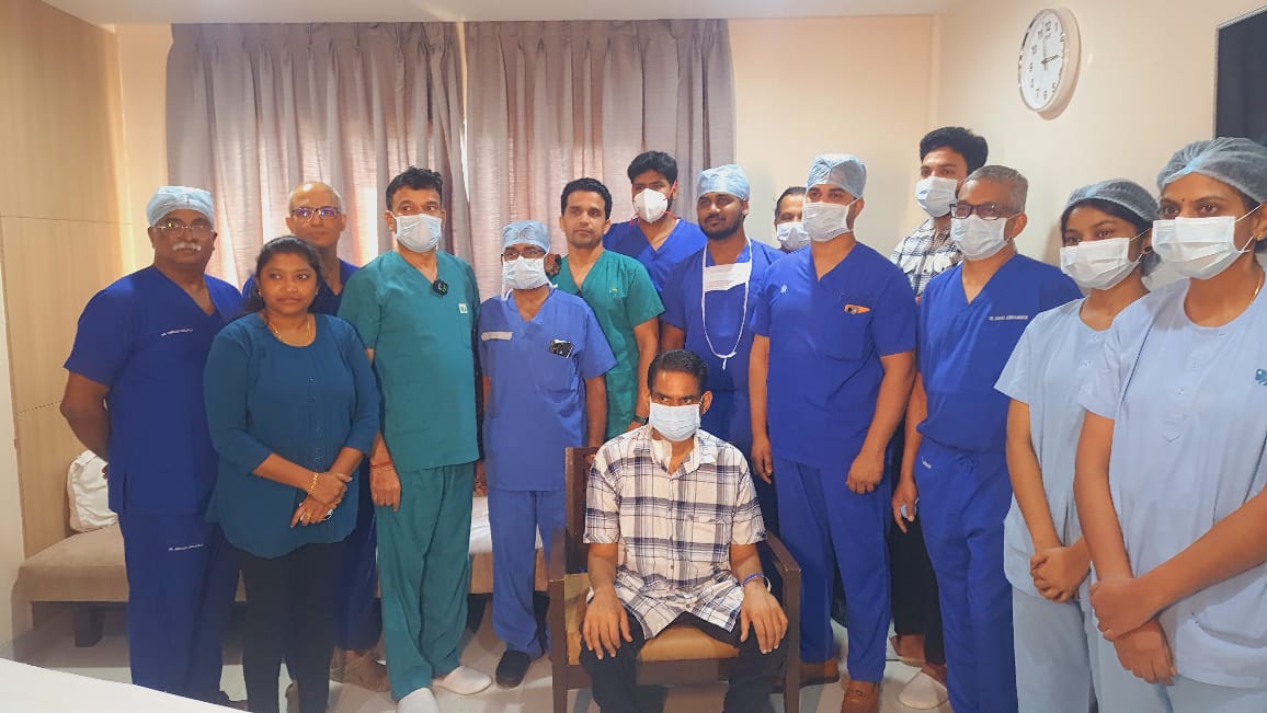
Chennai, April 22nd, 2025: MGM Healthcare successfully performed a complex heart surgery on a 6–year–old boy who was suffering from severe Left Ventricular Outflow Tract (LVOT) obstruction, a critical condition that severely blocked blood flow from his heart to the rest of his body in a dextrocardia heart. The medical team opted for the ‘Modified Konno Procedure’, a highly specialised procedure that relieves the obstruction while still preserving the child’s native heart valves – instead of conventional procedures like Ross procedure that would have involved implantation of artificial valves. The young patient has recovered well, with improved heart function.

The patient had been experiencing chest pain and shortness of breath. Upon his admission, the medical team promptly diagnosed his condition of severe LVOT obstruction that could lead to heart failure or other life-threatening complications, if left untreated. Recognising the urgency, the hospital carried out the surgical intervention to relieve the blockage and restore normal blood flow from the heart. This life saving surgery was performed at MGM Healthcare in collaboration with Aishwarya Trust, ensuring the child receive world class care.
The surgery was successfully performed by Dr. R.K.R. Noveen Davidson, Senior Consultant, Pediatric Cardiac Surgery, at MGM Healthcare with anesthesia support provided by Dr. Suresh Rao K.G and pediatric cardiology team.
Talking about the procedure, Dr. Noveen Davidson said that LVOT is a serious heart condition in which the flow of blood from the left ventricle, the main pumping chamber of the heart, to the rest of the body is significantly blocked. To treat the obstruction a Modified Konno Procedure, a complex and advanced heart surgery, was chosen to enlarge the narrowed outflow tract of the left ventricle. The procedure preserves the native valves, thus minimising the risk of repeated valve replacement surgeries in the future, as is the case with conventional Ross procedure.
“The operation was done through the chest bone, with the patient connected to a heart-lung machine to maintain circulation during surgery. The boy’s body was also cooled to a safe, controlled temperature to protect vital organs during surgery. After carefully opening the heart, we identified and removed thickened tissue and fibrous bands that were causing the blockage below the aortic valve. Since the outflow tract was still narrow, we made an additional opening through the right side of the heart to further clear the pathway. We used a medical-grade patch (synthetic patch – PTFE) to widen the passage and enlarged the aortic valve using a patch made from the patient’s own heart covering (pericardium). The heart was gradually brought back to normal temperature and rhythm and began beating on its own. A heart scan during surgery confirmed that the valve was working well and the blood flow from the heart had greatly improved, with no gradient. The patient was safely taken off the heart-lung machine and moved to recovery in stable condition,” he added.
Further Dr. Noveen Davidson added, “Modified Konno Procedure is especially beneficial in patients with severe narrowing that cannot be managed with conventional techniques. It helps improve blood flow from the heart to the rest of the body, reduces strain on the heart muscle, and significantly enhances the patient’s quality of life and long-term heart function”.
IDEA® World Convention in Sacramento, California: July 17-19, 2025
IDEA® World Convention in Sacramento, California: July 17-19, 2025
Health & Fitness Leaders from 80 Countries Converge to
‘Inspire the World to Fitness®’
SAN DIEGO, California – For the first time since its 1982 inception, the IDEA® Health and Fitness Association will be hosting fitness, health, nutrition, and wellness professionals, educators, and researchers representing over 80 countries worldwide, in California’s Capitol City of Sacramento, for the 2025 IDEA® World Convention July 17-19, 2025:
- 2025 IDEA® WORLD: https://www.ideafit.com/fitness-conferences/2025-idea-world/
- IDEA CEO Amy Thompson Announcing 2025 IDEA® WORLD in Sacramento: https://bit.ly/IDEACEOSacramento
- IDEA World Speakers & Schedule: https://bit.ly/IDEAWorld2025Brochure
More than a three-day educational event — IDEA® World is a dynamic, annual celebration of wellness and inspiration — crucial for personal trainers, group fitness instructors, yoga mind-body leaders, business owners, researchers, educators, nutritionists and dieticians, sports medicine and physical therapists, and any/all healthy body and mind advocates.
Featuring over 175 sessions presented by the most respected, global health and fitness leaders, IDEA® World provides and inspires attendees to advance their professional skillset and certifications, earn 20+ continuing education credits (CECs), elevate and enhance their network, and learn/adopt many of the industry’s latest research, developments, training, equipment, and tools. Among the educational tracks include:
- Personal Trainer Techniques
- Nutrition & Behavioral Change
- Group Exercise
- Mind-Body
- Fitness Business & Professional Development
- Exercise Science
- Special Populations (such as “Active Aging”)
Also – open and FREE to the public – will be the “IDEA World Fitness & Nutrition Expo”, featuring a diverse array of exhibitors and sponsors (such as Lifetime Fitness, WeGym, Keiser, Peeler’s Apparel, Perform Better, Merrithew and the National Academy of Sports Medicine, among others):
Hear directly from leading brands on why sponsoring or exhibiting at the 2025 IDEA® World Fitness & Nutrition Expo continues to deliver unmatched value-watch the video to see how these partnerships are driving real business growth. https://bit.ly/IDEAExhibitors.
Featuring live demonstrations and product sampling, the Expo will include exhibitors from workout and wellness equipment; fit-tech and nutrition brands; and group fitness, apparel, education, and business services; among others. For more information about sponsorship and exhibitor space availability and attendee demographics: https://bit.ly/IDEAWorldProspectus.
2025 IDEA® World attendees will also enjoy networking and special events, such as the “Golden Gala of Distinction” and IDEA Fitness Awards (featuring 99-year-old Fitness Icon Elaine LaLanne presenting the “Jack & Elaine LaLanne Lifetime Achievement Award”).
“Our IDEA team is very excited to host 2025 IDEA® World in Sacramento this year,” states Amy Thompson, CEO of the IDEA Health and Fitness Association. “We selected Sacramento for its beauty, diversity, affordability, and easy access to the world’s most innovative, wellness programs, and popular recreation leaders. And we are designing our 2025 IDEA® World program to offer cutting-edge insights, motivational experiences, and a supportive community to fuel the journey of anyone – professional or personal – that’s interested in ‘inspiring the world to fitness!”
IDEA® World will be in the Sacramento Convention Center, North Hall, 1401 K St, Sacramento, CA 95814, on July 17-19, 2025. To register, exhibit or for more information: https://www.ideafit.com/fitness-conferences/2025-idea-world/.
Redcliffe Labs Diagnoses India’s First Case of DPH2-Related Diphthamide Deficiency Syndrome-2
22nd April 2025: Redcliffe Labs, a purpose-driven pan-India omnichannel diagnostics service provider, has diagnosed the first known case of DPH2–related diphthamide deficiency syndrome–2 in India. This groundbreaking diagnosis has been published in the prestigious Wiley American Journal of Medical Genetics Part A, marking a significant milestone in the field of medical genetics and broadening the clinical understanding of this rare genetic condition. The diagnosis was carried out by Dr. Vykunta Raju K. N from Indira Gandhi Hospital, Bangalore, and Interpretation was done by Dr Himani Pandey, Lab Head of Genomics at Redcliffe Labs, and her team in collaboration with experts from leading medical institutions.
Identifying the DPH2 gene mutation enables healthcare providers to consider genetic counselling for families with a history of consanguinity. Early detection of this rare genetic disorder can facilitate timely intervention and management, potentially improving the quality of life for affected individuals. The ability to detect such mutations also aids in formulating more targeted and precise therapeutic strategies.
As per the diagnosis, the patient is a 1-year-9-month-old female who presented with a complex set of symptoms, including developmental delay, short stature, seizures, sparse hypopigmented hair, and dysmorphic facial features. Uniquely, the child also exhibited epileptic seizures, behavioural abnormalities, and neuroimaging findings such as cerebral atrophy and white matter hyperintensities – features not previously reported in other cases of this syndrome. The clinical picture also included developmental regression, autistic features, and hypotonia, making it a distinct and complex presentation.
The diagnosis was confirmed through whole-exome sequencing, which revealed a pathogenic nonsense variant in the DPH2 gene. This mutation truncates the DPH2 protein’s C-terminal, likely impacting its role in protein synthesis. According to the American College of Medical Genetics and Genomics (ACMG) standards, the identified variant has been classified as pathogenic. This is the first confirmed case of this syndrome in India and only the third reported case worldwide.
The patient exhibited delayed developmental milestones, including late attainment of neck control and the ability to sit with support, along with poor weight gain and feeding difficulties. Multiple episodes of generalized tonic seizures and epileptic spasms were documented, beginning at six months of age. Neuroimaging revealed cerebral atrophy, periventricular white matter hyperintensities, and prominent subarachnoid spaces, indicating significant neurological involvement. Additionally, the child showed distinct dysmorphic features such as a broad forehead with a high anterior hairline, sparse scalp hair, esotropia, a depressed nasal bridge, a bulbous nose, low-set ears, and brachydactyly in both hands and feet. This diagnosis holds immense significance for the medical community as it opens new avenues for preventive healthcare and prenatal diagnosis.
Dr. Himani Pandey, Lab Head Genomics of Redcliffe Labs said ,”This case exemplifies how integrative genomic approaches can elucidate the underlying molecular etiology of rare and clinically complex syndromes. Identifying a pathogenic DPH2 variant in an Indian patient not only contributes to the global repository of rare disease mutations but also enables more precise genotype-phenotype correlation. Such discoveries are vital to advancing personalized medicine, particularly in populations with high rates of consanguinity where recessive disorders often go undiagnosed.”
This discovery not only expands the known phenotypic spectrum of DPH2–related diphthamide deficiency syndrome–2 but also emphasizes the importance of including DPH2 mutations in diagnostic panels when evaluating developmental delays and epilepsy, particularly in populations with consanguineous backgrounds. Identifying this gene variant also paves the way for targeted research into the role of DPH2 in protein synthesis, potentially aiding in the development of therapeutic interventions.
Aditya, CEO & Founder of Redcliffe Labs, said, “This diagnosis reflects our commitment to leveraging advanced genomic technologies to uncover rare and complex genetic disorders. Identifying the DPH2–related diphthamide deficiency syndrome–2 in India not only highlights the capabilities of our team but also sets a precedent for integrating precision genetics into routine diagnostics. Our focus remains on making genetic insights accessible and impactful for patient care.”
Redcliffe Labs remains committed to pioneering advancements in genetic diagnostics and fostering research collaborations to improve patient outcomes. As a leading diagnostic and molecular genetics laboratory in India, they continue to deliver innovative healthcare solutions to address complex medical challenges.
Indoor Air Pollution: A Silent Trigger for Cancer Risk
Dr. Boppana Sai Madhuri, Consultant, Medical Oncologist, HCG Cancer Center – Vijayawada
As we go about our daily lives, it’s easy to overlook the air we breathe indoors. However, the truth is that indoor air quality plays a significant role in our overall health, particularly when it comes to cancer risk.
The Indoor Air Pollution Problem
Indoor air pollution is a pervasive issue that affects millions of people worldwide. The air inside our homes, offices, and other buildings can be up to five times more polluted than the air outside. This is due to the presence of various pollutants, including volatile organic compounds (VOCs), particulate matter (PM), and radon. These pollutants can emanate from a range of sources, including building materials, furniture, household cleaning products, and heating systems.
The Link to Cancer Risk
Exposure to indoor air pollutants has been linked to an increased risk of cancer. The International Agency for Research on Cancer (IARC) has classified some indoor air pollutants, such as radon and PM, as carcinogenic to humans. Radon, a radioactive gas that can seep into buildings from the soil, is a known cause of lung cancer. Similarly, exposure to PM, which can come from sources like cooking and heating, has been linked to an increased risk of lung cancer and other respiratory diseases.
Other Indoor Air Pollutants and Cancer Risk
In addition to radon and PM, other indoor air pollutants have also been linked to cancer risk. For example, VOCs, which can be emitted by building materials, furniture, and household cleaning products, have been shown to cause cancer in animals. Similarly, exposure to secondhand smoke, which can linger in indoor air, has been linked to an increased risk of lung cancer and other respiratory diseases.
Reducing Cancer Risk through Improved Indoor Air Quality
While the link between indoor air quality and cancer risk is alarming, there are steps we can take to reduce our exposure to indoor air pollutants. Here are some strategies for improving indoor air quality:
- Use ventilation systems : Installing ventilation systems that exchange indoor air with fresh outdoor air can help reduce pollutant levels.
- Remove sources of pollution : Identify and remove sources of pollution, such as radon and secondhand smoke.
- Use air purifiers : Air purifiers can help remove pollutants from the air.
- Choose low-VOC products : Opt for building materials, furniture, and household cleaning products that emit low levels of VOCs.
The impact of indoor air quality on cancer risk is a serious concern that deserves our attention. By understanding the sources of indoor air pollution and taking steps to reduce our exposure, we can mitigate this hidden danger and create healthier indoor environments. The air we breathe indoors matters, and it’s up to us to take action to protect our health.
IVF Over the Age of 35 Still Offers Hope for Motherhood
Dr. Neelam Banerjee, Senior Consultant and Head of Department – IVF, Yatharth Hospital, Greater Noida
In today’s world, more women are choosing to have children later in life. Whether it’s due to career priorities, financial planning, late marriages, or personal reasons, the average age of starting a family is steadily rising, especially in urban India. But as the age increases, so do the concerns about fertility. One of the most common questions women over 35 ask is: “Will IVF work for me?” Unfortunately, this question is often surrounded by confusion, myths, and fear.
Understanding IVF and Age
After 30, the number and quality of a woman’s eggs start to reduce, and the drop becomes more noticeable after 35. By the time a woman crosses 40, natural conception becomes significantly more difficult. This is where assisted reproductive technologies like IVF (In-Vitro Fertilization) become relevant.
But here’s the good news, IVF can still be highly successful in women over 35. With the right medical care and timely intervention, thousands of Indian women in their late 30s and early 40s have gone on to have healthy pregnancies and babies through IVF.
Busting the Myths
Many people believe IVF doesn’t work after 35, but that’s simply not true. While it’s a fact that success rates are higher for younger women, many women in their late 30s and even 40s have conceived successfully with IVF. The outcome depends on several factors like the woman’s egg reserve, hormone levels, overall health, and the experience of the fertility clinic. Starting the process early and following medical guidance can make a big difference.
Another common myth is that women over 35 always need donor eggs. While donor eggs can be an option for women with very low egg quality, it’s not the rule. Many women in this age group are still able to conceive using their own eggs. Tests like AMH and ultrasound help doctors evaluate the chances and suggest the right path forward.
It’s also important to understand that IVF is not a magic solution. It increases your chances of getting pregnant but does not guarantee a baby. Sometimes, it takes more than one cycle to succeed. Success depends on many things – the woman’s age, the health of the sperm, how well the embryo develops, and the condition of the uterus. Thankfully, advances in IVF technology have helped improve success rates, especially for older women.
IVF in India
India has seen a sharp rise in the number of couples seeking IVF treatment in the past decade. A study by the Indian Society of Assisted Reproduction, shows that over 25% of IVF patients are now above the age of 35, with urban areas seeing higher demand. The awareness is slowly increasing, but more needs to be done to educate women that fertility is not something to be taken for granted.
Taking the Right Steps
If you are over 35 and trying to conceive for more than 6–12 months without success, don’t delay seeking help. A simple fertility workup, including AMH test, ultrasound, and hormone levels can give a clear picture of where you stand.
IVF after 35 is not a dead-end, it’s a carefully managed medical path that has helped countless women become mothers. While age is a factor, it is not the only one. With growing awareness, access to better medical care, and open conversations, more women can make informed choices about their fertility.
Navigating Motherhood with Occupational Therapy

By: Dr. Joseph Sunny Kunnassery- Founder of Prayatna, Kochi
Becoming a mother is one of life’s most beautiful and most challenging transformations. It isn’t just a new chapter,it’s a whole new book.From the first thought of starting a family to the reality of sleepless nights and diaper duty, the journey through pregnancy, birth, and early parenthood can feel like an emotional rollercoaster. Amid the flood of hormones, endless check-ups, and well-meaning advice, there’s one healthcare profession often overlooked yet quietly making a world of difference- Occupational Therapy.
Occupational Therapy is about helping people do the things that occupy their day,things that give life meaning. And during pregnancy and early parenthood, those “occupations” can be something as simple (and as overwhelming) as changing a diaper, powering through sleepless nights, figuring out how to shower with a newborn in the house, or navigating the emotional rollercoaster that comes with being someone’s everything.
Though largely overlooked in maternity care, occupational therapists are uniquely positioned to support women at every stage of this life-changing transition. Whether helping manage morning sickness and mental fatigue during pregnancy, guiding a safe and dignified birth experience, or building healthy routines and bonds with a newborn, occupational therapy brings a holistic and empowering approach to maternal health and wellbeing.
OT Through the Journey
Even before pregnancy, occupational therapists play a proactive role in supporting women especially those with pre-existing physical or mental health conditions. The preconception phase can be filled with both hope and uncertainty, and occupational therapists work closely with individuals or couples to consider how existing health issues might affect pregnancy, childbirth, and the experience of becoming a parent. For those undergoing fertility treatments like IVF, occupational therapy offers emotional and lifestyle support, helping prospective parents navigate stress, adjust routines, and prepare their environment for the changes ahead.
As pregnancy progresses, occupational therapy becomes even more essential. This phase is not just about physical changes rather it involves a profound reconfiguration of daily life, roles, and routines. Expectant mothers may experience fatigue, aches, and a sense of being overwhelmed. Occupational therapists provide support in managing these physical symptoms while also helping women prepare for their shifting identities. They assist in modifying home environments for increased comfort and accessibility, suggesting ergonomic changes at work, and helping build flexible routines that balance rest, activity, and self-care. Their guidance empowers women to maintain a sense of control and well-being during a time of constant change.
When it comes time to give birth, occupational therapists continue to offer crucial, though often unrecognized, support. For women with disabilities, injuries, or health complexities, they ensure that the necessary equipment and support systems are in place to make the delivery process as safe, dignified, and empowering as possible.
But perhaps the postpartum period, often referred to as the fourth trimester, is where occupational therapy’s impact becomes most profound. It’s a time of immense transition, not just physically but emotionally and socially as well. While the spotlight tends to shift to the newborn, new mothers often face sleep deprivation, identity shifts, physical recovery, and mental health challenges. Occupational therapists work with mothers to support what are called co-occupations-activities that both the mother and baby engage in together, such as feeding, sleeping, and playing.
This way, occupational therapists help mothers regain confidence and competence in their new roles. They offer breastfeeding support, guidance in newborn care, pain management strategies, and help in setting up home routines that allow space for rest, bonding, and self-nurturing. They also recognize the signs of postpartum mood and anxiety disorders and provide early interventions, or collaborate with mental health professionals when needed and thereby provide holistic care that respects the mother’s physical health, emotional state, and personal environment.
As the first year of parenthood unfolds, occupational therapists remain vital companions in helping families find their balance. They support mothers returning to work, help create routines that actually work, and offer realistic ways to reintroduce exercise, hobbies, rest and support the ongoing development of the child.
Occupational therapists also bring a mental health perspective to their practice, with training that includes the recognition of trauma, anxiety, and depression. This allows them to provide tailored coping strategies that are practical and sustainable. They may lead therapeutic groups, connect families with community resources, or simply provide a space where a new mother can be heard and supported.
As the saying goes, when a baby is born, so is a mother. And yet, amidst the joy and chaos of new life, mothers often find themselves lost in the chaos. Occupational therapy helps them find their footing again not by prescribing a rigid formula for parenting, but by helping each woman build her own personal manual, helping mothers not just survive, but thrive.
India Leads in Liver Disease — ‘Food is Medicine’ Marks World Liver Day 2025
Mumbai, April 19, 2025 – On World Liver Day 2025, the global health community is turning its focus to a critical yet often overlooked issue: liver health. This year’s theme, “Food is Medicine,” highlights the powerful link between nutrition and liver function. In India—where liver disease has reached alarming levels—the message carries even greater urgency.
World Liver Day is observed every year on April 19 to raise awareness about liver health and promote prevention strategies. With liver-related deaths rising worldwide, the emphasis on food as a tool for prevention and healing is both a wake-up call and a practical solution. According to current estimates, India records approximately 268,580 liver disease deaths each year, accounting for 3.17% of all deaths in the country. More strikingly, this represents 18.3% of global liver-related deaths, making India the highest contributor to liver disease fatalities worldwide.
Commenting on the situation, Dr. Aditya Verma, Consultant Gastroenterologist at Wockhardt Hospitals, Mira Road, said: “India is facing a silent epidemic of liver disease, and much of it is driven by what we eat. Everyday food choices can either fuel liver damage or support healing.” The leading causes of liver disease in India include fatty liver disease , hepatitis infections, alcohol-related liver damage, and lifestyle-related metabolic conditions. A major contributing factor is a poor diet—often high in processed foods, sugar, and unhealthy fats.
This year’s World Liver Day theme promotes simple, sustainable dietary changes, such as:
- Reducing intake of refined sugars, white flour, and fried foods
- Eating more whole grains, vegetables, fruits, and legumes
- Choosing healthy fats like those in nuts, seeds, and olive oil
- Staying well-hydrated and limiting alcohol consumption
Dr. Aditya Verma, Consultant Gastroenterologist added, “Food is our first and most effective medicine. By shifting to a balanced, nutrient-rich diet, people can reduce their risk of liver disease—and in many cases, even reverse early-stage damage.” As liver diseases continue to rise across the country, medical experts are urging individuals to take control of their health through mindful eating. World Liver Day 2025 serves as a reminder that prevention often begins on our plates.
A Wearable Smart Insole Can Track How You Walk, Run and Stand
Newswise — COLUMBUS, Ohio – A new smart insole system that monitors how people walk in real time could help users improve posture and provide early warnings for conditions from plantar fasciitis to Parkinson’s disease.
Constructed using 22 small pressure sensors and fueled by small solar panels on the tops of shoes, the system offers real-time health tracking based on how a person walks, a biomechanical process that is as unique as a human fingerprint.
This complex personal health data can then be transmitted via Bluetooth to a smartphone for quick and detailed analysis, said Jinghua Li, co-author of the study and an assistant professor of materials science and engineering at The Ohio State University.
“Our bodies carry lots of useful information that we’re not even aware of,” said Li. “These statuses also change over time, so it’s our goal to use electronics to extract and decode those signals to encourage better self health care checks.”
It’s estimated that at least 7% of Americans suffer from ambulatory difficulties, activities that include walking, running or climbing stairs. While efforts to manufacture a wearable insole-based pressure system have risen in popularity in recent years, many previous prototypes were met with low energy limitations and unstable performances.
To overcome the challenges of their precursors, Li and Qi Wang, the lead author of the study and a current PhD student in materials science and engineering at Ohio State, sought to ensure that their wearable is durable, has a high degree of precision when collecting and analyzing data, and can provide consistent and reliable power, said Li.
“Our device is innovative in terms of high resolution, spatial sensing, self-powering capability, and its ability to combine with machine learning algorithms,” she said. “So we feel like this research can go further based on the pioneering successes of this field.”
The study was recently published in the journal Science Advances.
This team’s system is also made unique through its use of AI. Using an advanced machine learning model, the wearable can recognize eight different motion states, including static ones like sitting and standing to more dynamic movements such as running and squatting.
Additionally, since the materials the insoles are made of are flexible and safe, the device, much like a smartwatch, is low-risk and safe for continuous use. For instance, after the solar cells convert sunlight to energy, that power is stored in tiny lithium batteries that don’t harm the user or affect daily activities.
Because of the distribution of sensors from toe to heel, the researchers could see how the pressure on parts of the foot is different in activities such as walking versus running.
During walking, pressure is applied sequentially from the heel to the toes, whereas during running, almost all sensors are subjected to pressure simultaneously. In addition, during walking, the pressure application time accounts for about half of the total time, while during running, it accounts for only about a quarter.
In health care, the smart insoles could support gait analysis to detect early abnormalities associated with foot pressure-related conditions (such as diabetic foot ulcers), musculoskeletal disorders (such as plantar fasciitis) and neurological conditions (such as Parkinson’s disease).
The new system also used machine learning to learn and classify different types of motion. That offers opportunities for personalized health management, including real-time posture correction, injury prevention and rehabilitation monitoring. Customized fitness training may also be a future use, the researchers said.
According to the study, these smart insoles showed no notable deterioration in performance after 180,000 cycles of compression and decompression, showing their long-term durability.
“The interface is flexible and quite thin, so even during repetitive deformation, it can remain functional,” said Li. “The combination of the software and hardware means it isn’t as limited.”
Researchers expect the technology will likely be available commercially within the next three to five years. Next steps to advance the work will be aimed at improving the system’s gesture recognition abilities, which, according to Li, will likely be helped with further testing on more diverse populations.
“We have so many variations among individuals, so demonstrating and training these fantastic capabilities on different populations is something we need to give further attention to,” said Li.
Bengaluru Hospital Saves 40-Year-Old Haryana Man with Heart Transplant
Bengaluru, 17th April 2025: A 40-year-old resident of Haryana, who had end stage heart failure, was given a new lease on life through a complicated heart transplant surgery performed at Apollo Hospital, Seshadripuram, Bengaluru.
The patient, who is a father of two and the sole breadwinner for his family, was diagnosed with Dilated Cardiomyopathy (DCM) with severe bi-ventricular dysfunction, a condition where both the left and right pumping chambers of the heart fail to function properly. His condition required frequent hospital admissions for recurrent heart failure, causing a rapid decline in his quality of life. After extensive treatments and worsening symptoms, he flew down to Bengaluru with the support of a specialized medical team to get the heart transplant done by Dr. Kumud Dhital, Program & Surgical Director for Heart & Lung at Apollo Seshadripuram.

Commenting on the patient’s condition while presented at the hospital, Dr. Kumud Dhital said, “Upon arrival at Apollo Hospitals, he showed severe symptoms of advanced heart failure. He required immediate intervention, including the insertion of an Intra-Aortic Balloon Pump (IABP) to stabilize his condition. Aggressive fluid management was performed to remove over 20 litres of excess fluid before he was deemed fit for a transplant.”
After a month-long wait, a suitable organ became available, and he underwent the heart transplant procedure, performed by Dr. Kumud Dhital with support from Senior Cardiothoracic & Vascular Surgeons Dr. Anand Subramaniyam Dr. Manoj Kumar & Dr. Prakash Ludhani, and anesthetists Dr. Pradeep Kumar and Dr. Srinivas Dhulipala. Owing to the concerted efforts of the multidisciplinary team, both the surgical procedure and post-operative recovery progressed smoothly. The patient was subsequently discharged within few weeks and returned to Sonipat, Haryana, to reunite with his family.
The patient’s brother expressed his gratitude, “It’s been an emotional journey, but seeing my brother healthy again is beyond words. We are deeply grateful to the donor family for giving my brother a second chance at life.”
While talking about the importance of organ donation, Dr. Kumud Dhital said, “this story highlights the transformative impact of organ donation on patients with end-stage organ failure. This patient’s survival was made possible by the selfless decision of a donor’s family and the coordinated efforts of donor coordination, organ registry, transplant teams, and other healthcare professionals. It also underscores the urgent need for greater awareness and participation in organ donation programs.”
Head of Apollo Hospitals Seshadripuram Mr. Uday Davda said “We Thank Jeevasarthakathe for their exceptional coordination in making organ donation a streamlined process in Karnataka. This is what had lead the state to be awarded the second-best state for organ donations in 2023 by the central government. We can restore many lives and families if the public at large come out to donate organs.”

